
 | George Kimpton & Viet Tran A middle aged male presents to the ED with a 48 hour history of chest pain & dyspnoea on a background of STEMI and a LVEF 50% in 2016. |
His chest pain is worse on inspiration and on lying flat.
Whilst in ED his pain is unremitting despite generous amounts of GTN.
- BP 89/50
- HR 105 SR
- Sat 90%RA
On auscultation he does not have any murmurs and scattered basal creps.
ECG is unchanged from previous records
POCUS is performed to challenge our pre-test probability of Sub/massive PE (by evidence of right heart strain) and/or pulmonary oedema:
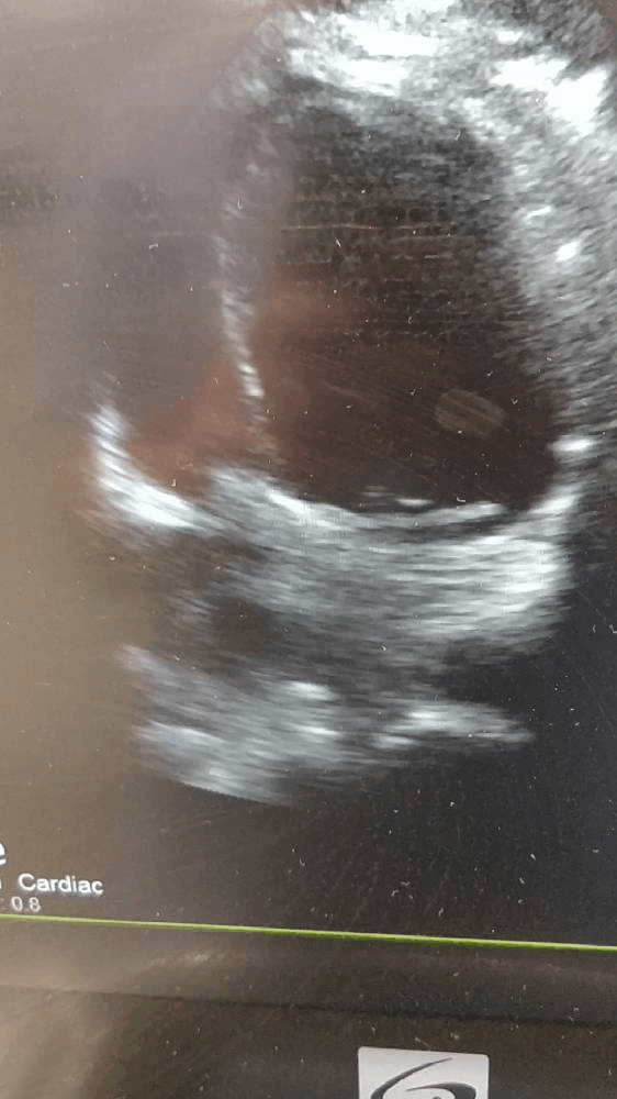
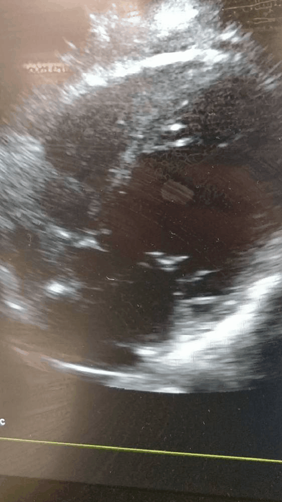

What do the above images show?
The above images consist of a lung field showing multiple B lines, and two cardiac views. These show a normally contracting, normally sized RIGHT ventricle without bowing of the septum towards the left ventricle in systole. The left ventricle appears large and dilated, with a visual EF of much less than the patient's prior stated EF of 50%.
The initial concern was for massive PE. The absence of echo features of right heart strain are reassuring. The presence of B lines is suggestive of fluid in the lung parenchyma - this is sensitive but specific. When added together with the history of PND, orthopneoa and the echo appearance of a hypokinetic left ventricle the clinical picture is suggestive of cardiogenic shock.
The initial concern was for massive PE. The absence of echo features of right heart strain are reassuring. The presence of B lines is suggestive of fluid in the lung parenchyma - this is sensitive but specific. When added together with the history of PND, orthopneoa and the echo appearance of a hypokinetic left ventricle the clinical picture is suggestive of cardiogenic shock.
What is the management for this patient?
With further investigative evidence of cardiac failure and ruling other causes of chest pain or precipitants of failure, he received IV furosemide 40mg BD.
A formal TTE the next day showed an EF of 40% with global hypokinesis and inferior wall hypokensis.
This gentleman was discharged home to his beloved alpaca the following day with a plan for an outpatient coronary angiogram.
A formal TTE the next day showed an EF of 40% with global hypokinesis and inferior wall hypokensis.
This gentleman was discharged home to his beloved alpaca the following day with a plan for an outpatient coronary angiogram.
 | George Kimpton is a first year emergency registrar with a background in academic geriatrics. He enjoys looking for B lines and sticking needles in people under ultrasound guidance. |
 RSS Feed